
Hand surgery care
A full range of services for the most common hand surgery complaints .
From the first consultation to post-operative follow-up, all in one hand - a transparent process, a personalised treatment plan. Below you will find a detailed description of the most common interventions.
1.
Consultation
Detailed anamnesis, targeted examination, clear explanation
2.
Diagnosis & Plan
Clear diagnosis, conservative or surgical options
3.
Treatment & follow-up
Intervention, control tests, rehabilitation plan
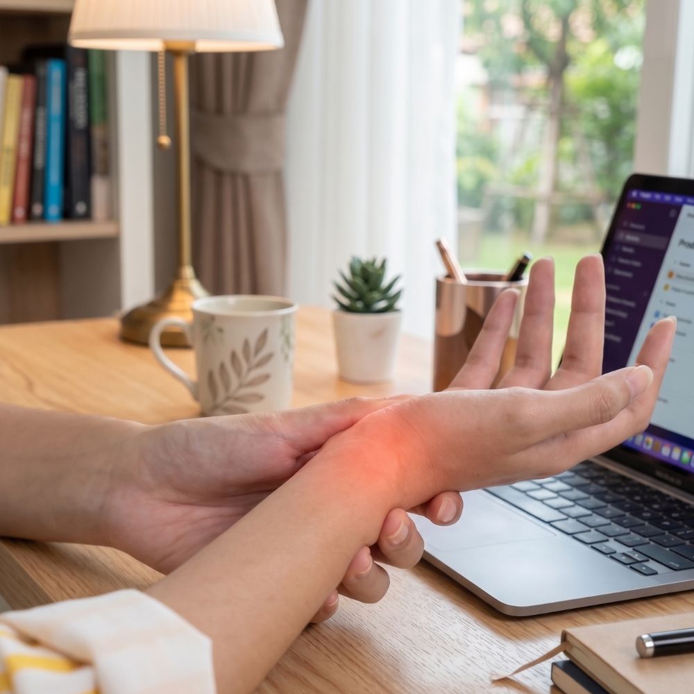
Carpal Tunnel Syndrome
Carpal tunnel syndrome is one of the most common nerve compression conditions of the hand. It occurs when the median nerve becomes compressed at the level of the wrist. This can cause numbness, tingling, pain or clumsiness in the hand. Symptoms most often affect the thumb, index finger, middle finger and part of the ring finger. Night-time numbness, the need to “shake out” the hand, and, in more advanced cases, reduced grip strength may also occur.
The aim of surgery is to release the compressed nerve. This can be performed either through a traditional open approach or by an endoscopic technique. In open surgery, the tight ligament is divided through an incision in the palm. In endoscopic surgery, the release is performed through a smaller incision using a camera. Both methods are effective. The choice of technique always depends on the symptoms, the anatomy and the findings of the personal consultation.
The aim of the operation is to relieve the pressure on the nerve. This can be done from conventional open-cut mining, or using endoscopic techniques In open surgery, the ligament is cut via an incision in the palm of the hand. In endoscopic surgery, the procedure can be performed via a smaller incision using a camera. Both methods are effective; the choice of technique is always based on the specific symptoms, anatomical conditions and a personal consultation.
During the consultation, we will discuss in detail whether surgery is necessary in your case, or whether conservative treatment is still a reasonable option. My aim is to give you a clear explanation of the cause of your symptoms, the available treatment options, and the advantages and limitations of both open and endoscopic surgery.
Not sure whether you need surgery?
Let us discuss it in person and choose the treatment option that is most appropriate for you.
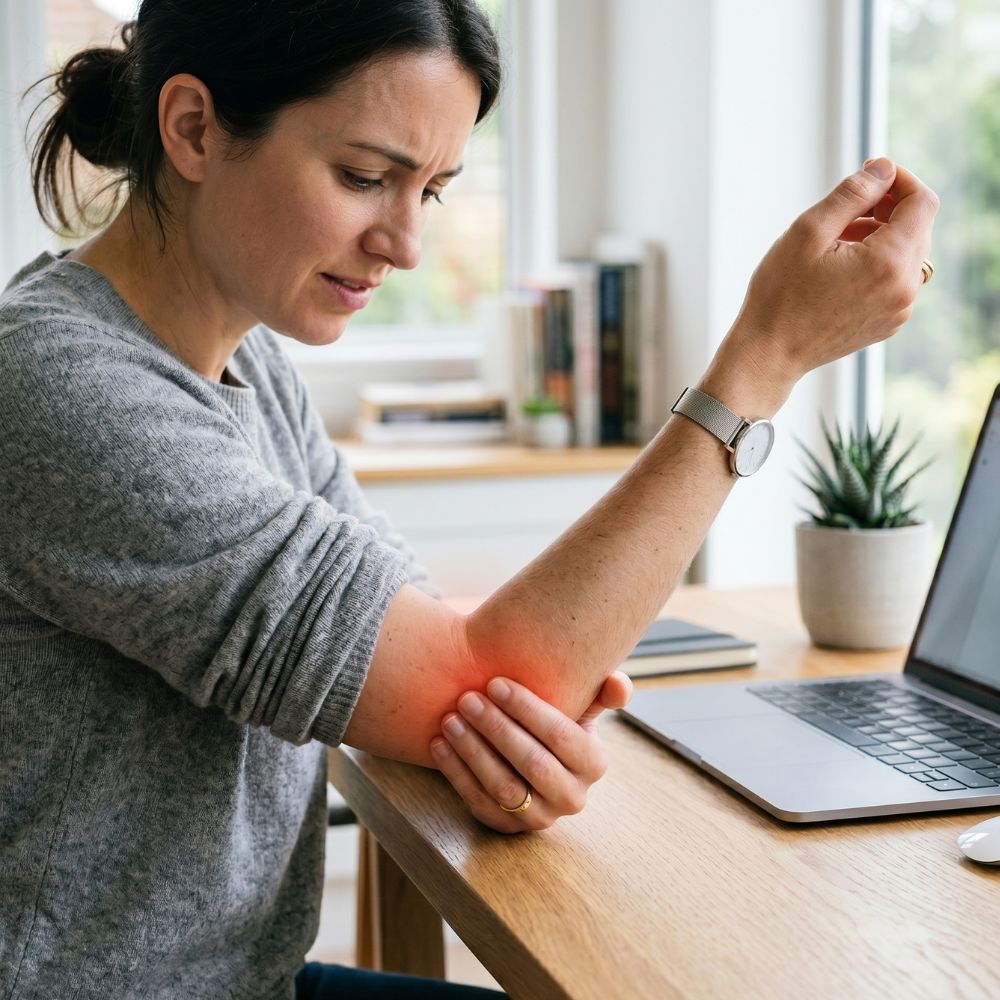
Cubital Tunnel Syndrome
Cubital tunnel syndrome is the second most common nerve compression condition. It affects the ulnar nerve, which runs along the inner side of the elbow. Compression of this nerve can cause numbness, tingling, pain or clumsiness, most often in the little finger and ring finger. In more advanced cases, grip strength may decrease, fine movements may become difficult, and muscle weakness or wasting may develop.
Symptoms often worsen when the elbow is bent, for example while using a phone, driving, sleeping or leaning on the elbow for a long time. Diagnosis is based on a detailed discussion of the symptoms and a physical examination of the hand and elbow. If needed, nerve conduction studies or ultrasound examination can help assess the degree of nerve involvement. In milder cases, non-surgical treatment may be considered, such as night-time elbow splinting, avoiding pressure on the elbow, anti-inflammatory treatment and targeted lifestyle advice.
If symptoms are persistent, worsening, or if numbness, loss of sensation or muscle weakness develops, surgical treatment may be required. The aim of the operation is to release the nerve. This can be done through a traditional open procedure, and in selected cases by an endoscopic technique. During open surgery, the nerve is released under direct vision and, if necessary, transposition of the nerve may also be considered. In endoscopic surgery, the nerve is released through a smaller incision using a camera by conventional open surgery, or, in certain cases using endoscopic techniques as well. In open surgery, the nerve is released via a direct incision, and nerve transposition may also be considered if necessary. In endoscopic surgery, the nerve can be released through a small incision using a camera.
Both surgical options have their role. The appropriate technique is always selected according to the symptoms, physical findings, nerve conduction results and anatomical factors. It is also important to assess whether the nerve remains stable during elbow flexion or whether it moves or “snaps” over the inner side of the elbow. During the consultation, we will discuss whether surgery is necessary in your case and, if so, which surgical option may be the most suitable.
Not sure whether you need surgery?
Let us discuss it in person. During the consultation, I will examine your symptoms in detail, clearly explain the possible causes, and recommend surgery only when it is truly indicated. In milder cases, conservative, non-surgical treatment can often be an appropriate solution.
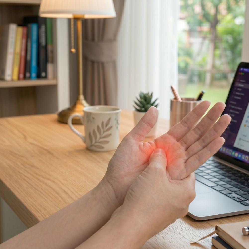
Trigger Finger
Trigger finger is a common tendon condition of the hand. The flexor tendon of the finger has difficulty gliding through a narrowed part of the tendon sheath. This can cause pain, catching or a characteristic “triggering” sensation during finger movement. The symptom is usually noticed during bending or straightening of the finger. In more severe cases, the finger may become locked and can only be straightened with assistance.
Diagnosis can usually be made based on the symptoms and a physical examination of the hand. In milder cases, the first treatment options may include rest, splinting, anti-inflammatory treatment or a steroid injection. The aim of the steroid injection is to reduce inflammation and swelling around the tendon sheath, thereby improving tendon gliding. In many cases, this can restore painless movement without surgery.
If symptoms persist, return or the finger regularly locks, surgical treatment may become necessary. The aim of the procedure is to release the A1 pulley so that the flexor tendon can move freely again.
A particularly useful option is percutaneous treatment without an incision. During this procedure, the narrowed part of the tendon sheath is released through a small puncture, without a larger skin incision. The advantage of the percutaneous technique is that the A1 pulley can be released without an open incision, which may allow faster rehabilitation. However, not every trigger finger is suitable for this method; the appropriate technique is always selected after a personal examination.
In some cases, open surgery is recommended. This involves releasing the A1 pulley through a small incision in the palm. Open surgery may be preferable when the anatomy, previous treatments, more pronounced symptoms or other factors make direct visualisation the safer and more precise option.
During the consultation, I will examine your symptoms in detail and we will discuss the non-surgical options, the role of steroid injection, and the advantages and limitations of both percutaneous and open surgery. My aim is to perform an intervention only when it is indicated and to choose the treatment option that is best suited to you.
Are you bothered by a finger that gets stuck or a painful catching/locking finger?
Let us discuss it in person. In many cases, an injection can provide significant improvement, while percutaneous or open surgical treatment is available when needed.
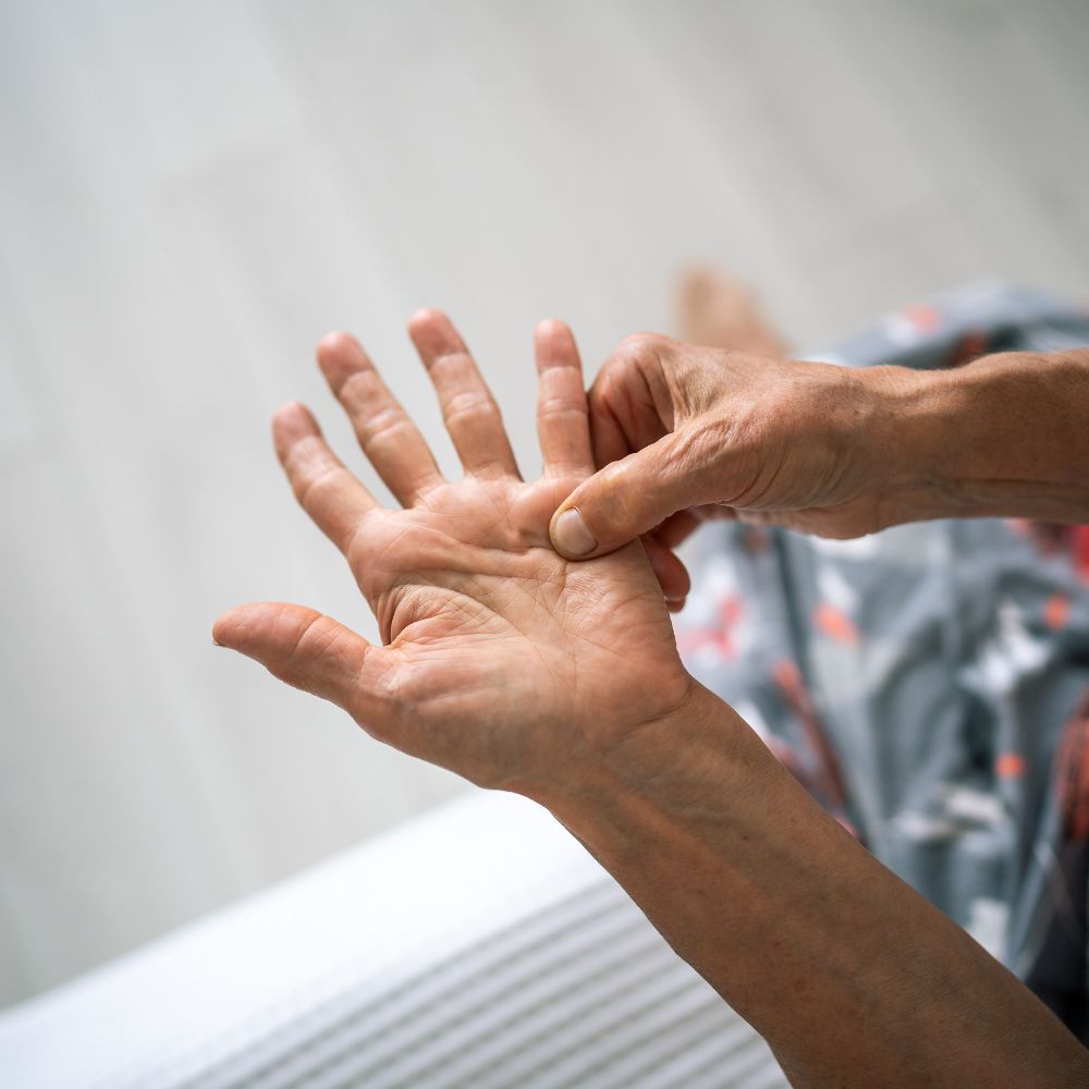
Dupuytren’s Contracture
Dupuytren’s contracture is a benign condition involving thickening and tightening of the connective tissue in the palm. Firm nodules and cords may appear in the palm, and over time these can pull the fingers into a bent position. The ring finger and little finger are most commonly affected, although other fingers may also be involved.
At first, the condition may present only as a palpable lump in the palm and does not necessarily cause pain. Later, straightening the fingers may become more difficult, the palm may no longer lie flat on a table, and hand function may become increasingly limited. It can become troublesome during activities such as shaking hands, putting on gloves, reaching into a pocket or doing sports.
Diagnosis is usually made by physical examination. During the consultation, I will examine the palmar cords, finger motion, the degree of contracture and the extent to which the condition affects everyday hand use. Not every Dupuytren’s change requires immediate intervention; in milder cases, regular follow-up and observation may be sufficient.
If the finger contracture causes functional problems, several treatment options may be considered. One less invasive procedure is percutaneous aponeurotomy, also known as needle aponeurotomy. During this procedure, the contracted palmar cord is divided through small punctures without a larger incision. Its advantages include a smaller intervention, potentially faster wound healing and a quicker return to everyday hand use. However, not every case is suitable for this method.
In more advanced, complex or recurrent cases, open surgery may be recommended. In this procedure, the thickened and contracted tissue is removed through an incision under direct vision. Open surgery can allow a more precise correction, especially when several fingers are affected, the contracture is severe or joint movement is significantly limited.
The most appropriate treatment is always chosen after a personal examination. The affected finger, the degree of contracture, the condition of the skin, previous treatments and the impact on everyday life must all be taken into account. The goal is not only to straighten the finger, but also to improve the function of the hand.
Is your finger bending inwards, or can you feel a lump in your palm?
Let us discuss it in person. During the consultation, I will assess whether observation is sufficient, or whether percutaneous aponeurotomy or open surgical treatment may be indicated.
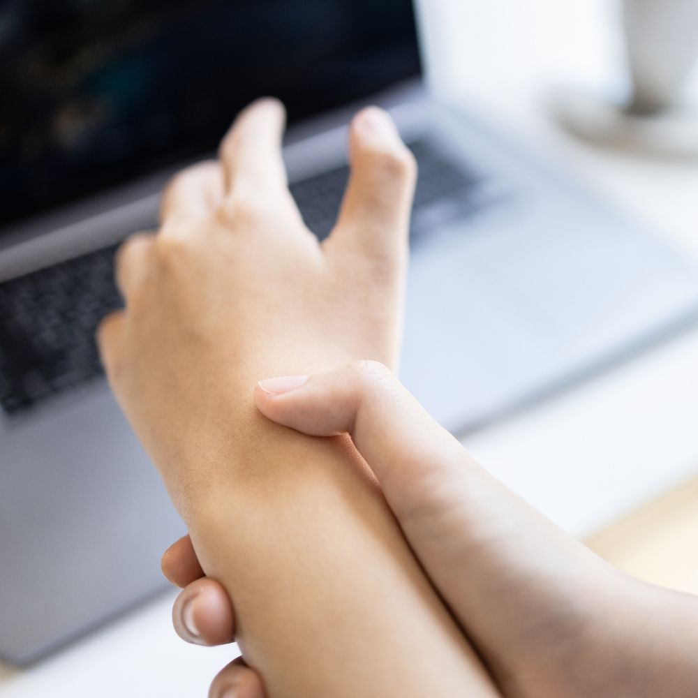
De Quervain’s Syndrome
De Quervain’s syndrome is a painful tendon sheath condition on the thumb side of the wrist. The tendons that move the thumb pass through a narrowed tendon sheath at the wrist, which can cause pain, swelling and limited movement. Symptoms are often aggravated by lifting, twisting movements, holding a small child, sports or repeated thumb use.
Diagnosis is usually based on the symptoms and physical examination. Tenderness on the thumb side of the wrist and pain during certain provocation tests are typical findings. Imaging is usually not required, but in uncertain cases ultrasound or other examinations may help confirm the diagnosis.
In milder cases, immobilisation may be recommended as a first step. A splint that rests the thumb and wrist can reduce the load on the tendons, thereby decreasing pain and inflammation. In addition to splinting, it may be important to avoid provoking movements, temporarily reduce the load on the hand and, if needed, use anti-inflammatory treatment.
For more persistent or more severe symptoms, a steroid injection can be given around the affected tendon sheath. The aim of the injection is to reduce inflammation and swelling and to help the tendons glide more freely. In many cases, this can lead to significant improvement without surgery, especially when combined with appropriate splinting and activity modification.
If symptoms persist or return despite conservative treatment, surgery may become necessary. The aim of the procedure is to release the narrowed tendon sheath so that the thumb tendons can glide freely again. The operation is usually performed through a small incision under local anaesthesia.
The choice of treatment is always based on a personal examination. The duration and severity of symptoms, the load placed on the hand, previous treatments and the degree to which pain limits everyday life all need to be considered. The goal is not simply to reduce pain, but to restore safe and reliable hand use.
Do you have pain on the thumb side of your wrist?
Let us discuss it in person. During the consultation, I will assess whether splinting or a steroid injection may be sufficient, or whether surgical release is indicated.
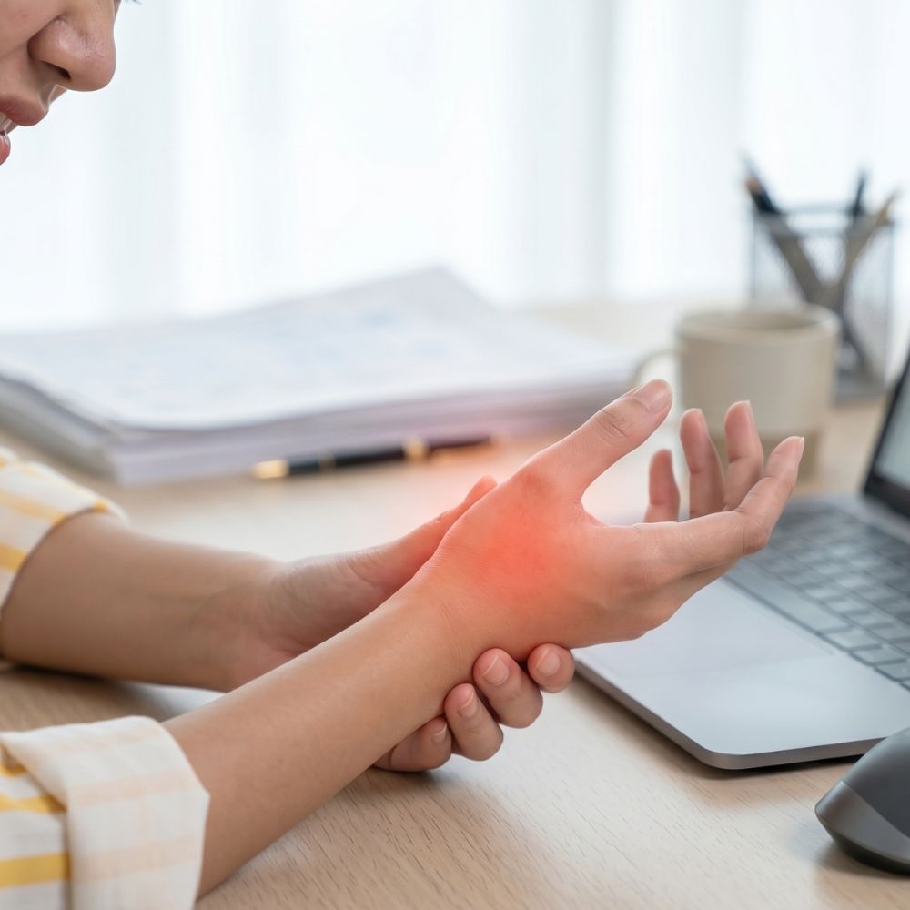
Thumb Basal Joint Arthritis
Osteoarthritis of the thumb basal joint, also known as thumb carpometacarpal (CMC) arthritis, is a common joint condition of the hand. In this condition, the cartilage surface at the base of the thumb gradually wears down, causing pain, difficulty with loading and restricted movement. Symptoms often appear when turning a key, opening a jar, performing twisting movements, writing or gripping firmly. In advanced cases, the base of the thumb may become enlarged or deformed, and pinch strength may decrease.
Diagnosis is based on a detailed discussion of the symptoms, examination of the hand and X-ray imaging. During the consultation, I will assess thumb motion, the location of pain, joint stability and the extent to which the condition limits everyday hand use.
In mild or moderate cases, non-surgical treatment is usually recommended first. An important part of this is immobilisation with a thumb and wrist splint that unloads the basal joint. Splinting can reduce pain, improve hand function and may help avoid or delay surgery. Anti-inflammatory treatment, targeted hand therapy and activity modification may also be considered.
If symptoms persist despite conservative treatment, if pain significantly limits hand use, or if the arthritis is advanced, surgical treatment may become necessary. One of the most commonly used procedures is trapeziectomy, in which the trapezium bone, one of the bones forming the arthritic basal joint, is removed. The aim is to eliminate painful bone-on-bone contact and improve thumb motion and load-bearing capacity. Trapeziectomy may be performed alone or combined with various stabilising or tendon reconstruction techniques.
After surgery, immobilisation and gradual rehabilitation are required. The exact surgical plan and aftercare always depend on the condition of the hand, the degree of arthritis, the patient’s lifestyle and the demands placed on the hand.
The most appropriate treatment is always selected after a personal examination. Not every case of thumb basal joint arthritis requires surgery: in many cases, splinting, activity modification and conservative treatment can reduce symptoms effectively. In advanced, painful cases, however, trapeziectomy can provide a long-term solution.
Do you have pain at the base of your thumb, or difficulty gripping, twisting or turning a key?
Let us discuss it in person. During the consultation, I will assess whether splinting and conservative treatment may be sufficient, or whether surgery, such as trapeziectomy, is indicated.
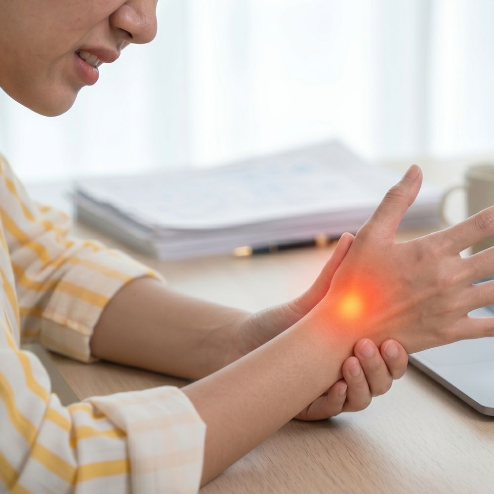
Pseudoarthrosis of the scaphoid
The scaphoid is one of the small but important bones in the wrist; fractures of this bone can be difficult to heal in certain cases. This is partly due to the bone’s unique blood supply and partly because the fracture often causes few symptoms initially, meaning it is often diagnosed late. If the fracture does not heal properly, The scaphoid is an important small bone in the wrist, and its fractures can sometimes be difficult to heal. This is partly due to the special blood supply of the bone and partly because scaphoid fractures may initially cause only mild symptoms, so they can be recognised late. If the fracture does not heal properly, a scaphoid non-union may develop. In this situation, persistent movement remains between the bone fragments, which can cause pain, difficulty with loading the wrist and, in the longer term, wrist arthritis may develop. In such cases, there remains persistent movement between the bone ends, which can cause pain, difficulty bearing weight and, in the long term, wear and tear of the wrist joint.
Symptoms often occur on the thumb side of the wrist. Pain may be felt when leaning on the hand, lifting, doing sports, performing push-ups or gripping strongly. Sometimes the original injury happened a long time earlier, and wrist pain only becomes persistent later.
Diagnosis is based on a discussion of the symptoms, physical examination of the wrist and imaging studies. In addition to X-rays, CT examination is often needed to assess the non-union accurately, including bone position, bone loss and possible deformity.
The choice of treatment always depends on the type of non-union, the location of the fracture, the condition and blood supply of the bone, and whether wrist arthritis is already present. In a true non-union, immobilisation alone is usually no longer sufficient.
In a symptomatic non-union, surgery is usually recommended. The aim is to improve the chance of scaphoid healing, restore bone alignment and reduce pain. During surgery, the non-union site is refreshed and a bone graft is placed into the missing or poorly healing area. The bone graft may be taken, for example, from the radius or ulna, and its purpose is to support new bone formation.
In addition to bone grafting, the scaphoid is usually fixed with a screw so that the bone fragments remain stable during healing. Stable screw fixation supports bone consolidation and helps the scaphoid heal in the correct position. In selected cases, a specialised vascularised bone graft may also be considered, especially when part of the scaphoid has a reduced blood supply.
After surgery, immobilisation, follow-up examinations and gradual rehabilitation are required. Healing can take several months, and the return to loading always depends on the pace of bone healing. The aim is to reduce pain, improve the load-bearing capacity of the wrist and reduce the risk of later arthritis.
Do you have persistent pain on the thumb side of the wrist after an old injury?
Let us discuss it in person. During the consultation, I will assess whether immobilisation and follow-up are sufficient, or whether surgery with bone grafting and screw fixation is indicated.
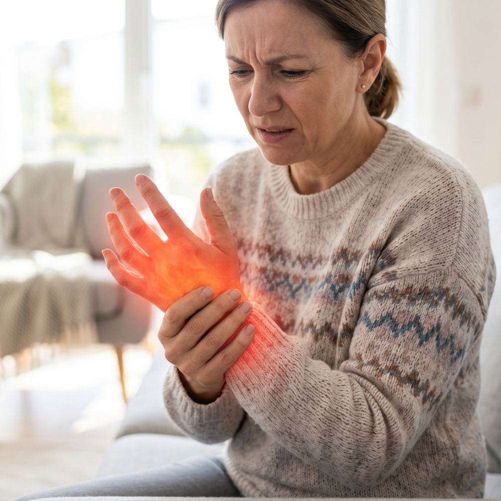
Sudeck’s Syndrome / Complex Regional Pain Syndrome
Sudeck’s syndrome, also known as complex regional pain syndrome (CRPS), is a condition that may develop after an injury or surgery. It is associated with persistent pain, swelling, stiffness and increased sensitivity. It often affects the wrist, hand or fingers, and the symptoms may be stronger than would be expected from the original injury alone.
The aim of treatment is to reduce pain, preserve hand motion and restore everyday hand use as early as possible. Treatment may include medication, physiotherapy, hand therapy, swelling control, desensitisation therapy and gradually increasing use of the hand.
In some cases, Sudeck-like symptoms may be maintained by nerve irritation or nerve compression. One example is irritative carpal tunnel syndrome, where increased sensitivity and compression symptoms of the median nerve contribute to ongoing complaints. In such cases, after appropriate assessment, surgical nerve release may also be considered. surgical decompression could also be an option.
Have persistent hand pain, swelling or stiffness developed after an injury or surgery?
Let us discuss it in person. During the consultation, I will assess whether conservative treatment, hand therapy or, in selected cases, surgery may be appropriate.
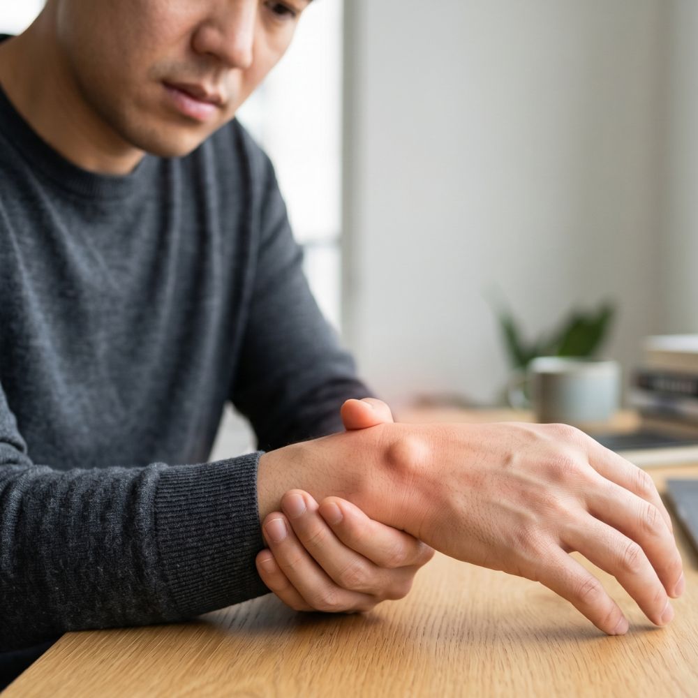
Ganglion
A ganglion is one of the most common benign conditions affecting the hand and wrist. It usually appears as a fluid-filled sac originating from a joint or tendon sheath. It can cause a palpable lump, tightness, cosmetic concerns, and occasionally pain or restricted movement.
The diagnosis can usually be made through a physical examination. In uncertain cases, an ultrasound scan, or less commonly an MRI scan, can help to determine whether it is indeed a ganglion and exactly where the lesion originates.
Not all ganglion cysts require surgery. In cases where there are no symptoms, simply monitoring the condition may be sufficient. If it causes pain, functional impairment or significant cosmetic concerns, aspiration or surgical removal may be considered. The procedure is usually performed under local or regional anaesthesia; in rarer cases, general anaesthesia may be required. The removed lesion may be sent for histopathological examination.
Can you feel a lump on your wrist or finger?
Let’s discuss this in person. During the consultation, I will assess whether observation is sufficient, whether imaging tests are required, or whether surgical removal of the ganglion is warranted.
Unsure about your complaint? Let's talk through!
It all starts with an accurate diagnosis. At the first consultation, we review the symptoms together and only move forward if warranted.